Hi everybody!! It’s me again!
Today, I would like to share with you guys how to perform Live/Dead Staining! This is a viability assay, similar to the process that Stanley has discussed in his post. However, this is not a quantitative assay.
I am using fluorescence to detect live / dead cells.
Concept
Live cells are capable of metabolism, therefore we add Calcein AM. Calcein is a fluorescent dye, and Calcein AM is the acetomethoxy derivate of calcein. Calcein AM is able to enter cells through intact cell membrane and thus is capable of determining live cells. Intracellular hydrolase enzymes (or intracellular esterases) will hydrolyze AM, and release green fluorescent anion calcein. This calcein will be retained in the cytoplasm of cells. Under fluorescence, a green cell is observed. On the other hand, when cells die, the plasma membranes of these cells are compromised. Hence AM will not be hydrolyzed. Instead, Ethidium homodimer-1 (EthD-1), is added. EthD-1 is also a fluorescent dye which will enter cells with compromised cell membrane. The dye will then bind to DNA in the nucleus. As EthD-1 is red in colour, it will give a red fluorescent under fluorescence.
We store them in 4oC. Since these are fluorescent dyes, they are sensitive to light. Hence when we add these chemicals to cells, we perform the experiment in the dark and cover in aluminium foil.
Calcein AM is diluted with cell culture medium/solution à 0.5ul/ml of solution (depending on what type of cells you using)
EthD-1 is diluted in cell culture medium/solution à1ul/ml of solution
Eg
- 1. Add 1ml of media to each well containing cells.
- 2. Add the dyes to cells. (Calcein AM: 0.5ul / EthD-1: 1ul)
- 3. Incubate at 37oC for 20-30 minutes.
- 4. Observe under fluorescent microscope.
Here 2 pictures for you guys to enjoy!
apparently blogger is down AGAIN, so it doesn't let me post pictures!
so here are the hyper- links instead.
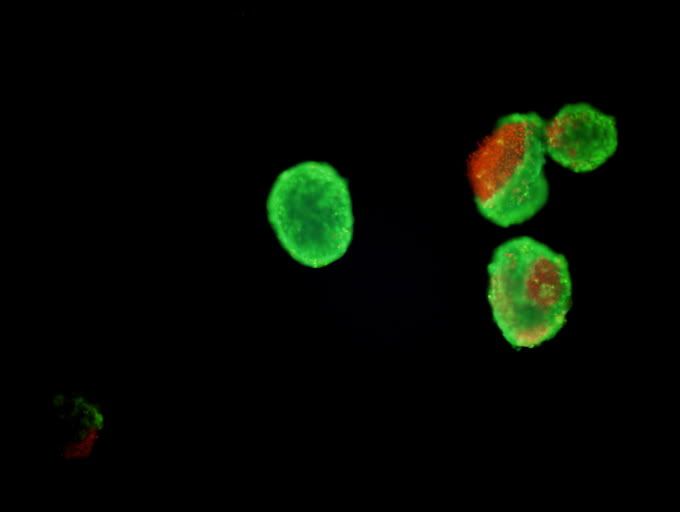
Figure 1: http://i201.photobucket.com/albums/aa29/sakura_1990/Image16.jpg
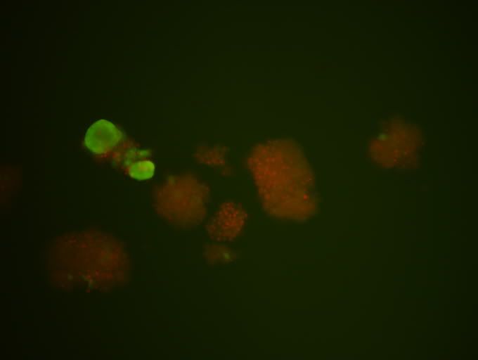
Figure 2: http://i201.photobucket.com/albums/aa29/sakura_1990/Image47.jpg
(here's the direct link if the above hyperlink doesn't work)
Figure 1 shows it all. There is live cells and going-to-die cells. As u can see from cell number 2 from the left, there is a portion of red fluorescent. This indicates that the cell membrane is compromised/ or cell has burst. This indicates that the cell is probably dead.
Figure 2: More obvious dead cells.
hopefully u guys can see the links. if there's any trouble, do leave a comment. I'll get back as soon as possible.
(the photos are released with permission).
posted by LIM JIA HUI (: tg01 0703605F