Hi everybody!! It’s me again!
Today, I would like to share with you guys how to perform Live/Dead Staining! This is a viability assay, similar to the process that Stanley has discussed in his post. However, this is not a quantitative assay.
I am using fluorescence to detect live / dead cells.
Concept
Live cells are capable of metabolism, therefore we add Calcein AM. Calcein is a fluorescent dye, and Calcein AM is the acetomethoxy derivate of calcein. Calcein AM is able to enter cells through intact cell membrane and thus is capable of determining live cells. Intracellular hydrolase enzymes (or intracellular esterases) will hydrolyze AM, and release green fluorescent anion calcein. This calcein will be retained in the cytoplasm of cells. Under fluorescence, a green cell is observed. On the other hand, when cells die, the plasma membranes of these cells are compromised. Hence AM will not be hydrolyzed. Instead, Ethidium homodimer-1 (EthD-1), is added. EthD-1 is also a fluorescent dye which will enter cells with compromised cell membrane. The dye will then bind to DNA in the nucleus. As EthD-1 is red in colour, it will give a red fluorescent under fluorescence.
We store them in 4oC. Since these are fluorescent dyes, they are sensitive to light. Hence when we add these chemicals to cells, we perform the experiment in the dark and cover in aluminium foil.
Calcein AM is diluted with cell culture medium/solution à 0.5ul/ml of solution (depending on what type of cells you using)
EthD-1 is diluted in cell culture medium/solution à1ul/ml of solution
Eg
- 1. Add 1ml of media to each well containing cells.
- 2. Add the dyes to cells. (Calcein AM: 0.5ul / EthD-1: 1ul)
- 3. Incubate at 37oC for 20-30 minutes.
- 4. Observe under fluorescent microscope.
Here 2 pictures for you guys to enjoy!
apparently blogger is down AGAIN, so it doesn't let me post pictures!
so here are the hyper- links instead.
Figure 1: http://i201.photobucket.com/albums/aa29/sakura_1990/Image16.jpg
Figure 2: http://i201.photobucket.com/albums/aa29/sakura_1990/Image47.jpg
(here's the direct link if the above hyperlink doesn't work)
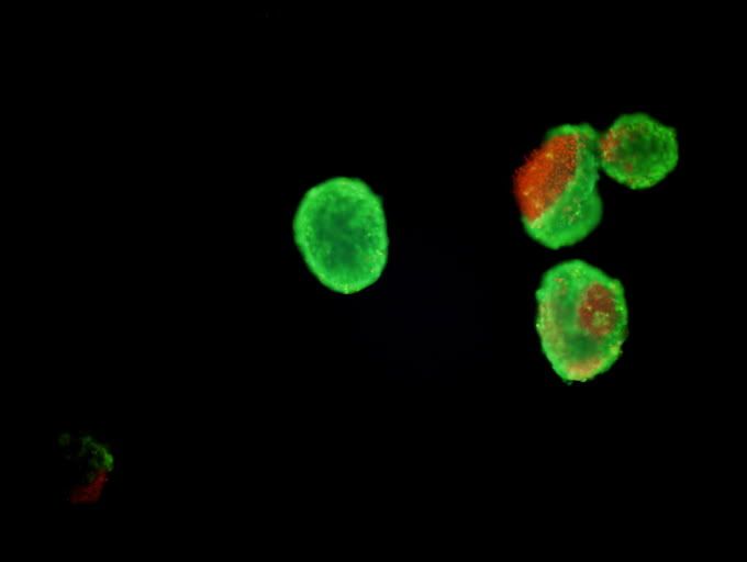
Figure 1 shows it all. There is live cells and going-to-die cells. As u can see from cell number 2 from the left, there is a portion of red fluorescent. This indicates that the cell membrane is compromised/ or cell has burst. This indicates that the cell is probably dead.
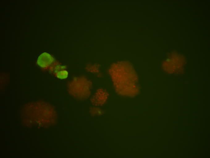
Figure 2: More obvious dead cells.
hopefully u guys can see the links. if there's any trouble, do leave a comment. I'll get back as soon as possible.
(the photos are released with permission).
posted by LIM JIA HUI (: tg01 0703605F
Hi joey,
ReplyDeleteI am just curious, you said that the EthD-1 will enter the dead cell membrane and bind just to the DNA inside the nucleus. If this is so, why is the dead cell in your figure 2,completely red. Is it due to the nucleus bursting and the discharge of the DNA contents to the whole cell?
Stanley
For image 1, there is a cell that appears green but with red fluorescent inside. So is it alive or dead?
ReplyDeleteAlvin
to stanley,
ReplyDeletethose cells are islets, if u read up on what islets are, they are actually islets of Langerhans found in pancreas, and inside each islet, there are numerous cells inside (including alpha/beta (just to name some)). so when the cell membrane burst, the contents will leak out. nucleus will also burst resulting in leakage. (:
to alvin,
these type of cells, we call it zombie-type cells(because it is semi-dead/alive. however, u can actually see that the cell contents are leaking out of cells, indicating that that particular cell is on the way to death. (so i would actually say that the cell is dead...)
jiahui (: hope this answers your questions!
hello Joey =D I like the picutres, very interesting to look at, ehheh
ReplyDeleteSo what will u do next yo? lolz. I mean since u said it's not a quantitative test, what's the purpose of viability staining assay? Is is just to see which cell is dead and which cell is alive? =D
Vo Thu Hong Anh [Jess]
0705364H
nopes, next I'll do MTT assays to determine the cell viability.
ReplyDeleteeg, treat cells with drug that kills the cells) --> perform live/dead staining (to observe if the cells really die,) --> perform MTT to determine cell viability --> can use the live/dead staining to confirm results of MTT
lim jia hui tgo1 0703605f
Jia Hui,
ReplyDeleteWhy do you perform both MTT and Calcein/EthD-1 staining?
Joey,
ReplyDeleteWhy cant trypan blue dye be used to differentiate between viable and nonviable cells - since this is a quantitative assay.
Li Yinliang Alex 0704894E
TG02 Group 8
9 September 2009
okay, thankz for the answer yo ^^ totally understood!
ReplyDeleteVo Thu Hong Anh [Jess]
0705364H
Typo: Not a quantitative assay
ReplyDeleteAlex.
Hey Joey
ReplyDeleteCorrect me if I'm wrong - I'm guessing that the EthD-1 is too big to pass through the nucleus of the living cells and hence we don't see the red fluorescence in the living cells?
Yvonee
0703189A
sorry for the late reply!
ReplyDeleteto dr lee,
MTT gives us the quantitative values of the total cell viability, while the live/dead staining gives us the visualization. eg, i have a total 100 cells. i pick 10 cells out for live/dead staining and the rest will be used for MTT assay.the live/dead staining will give a rough indication of cell viability while the MTT gives a confirmed indication.
to yvonee,
viable cells have an intact cell membrane, meaning the cell membrane is working to "select" what goes in and out of the cell. so, when cells are alive, the cell membrane dont allow ethd-1 to enter cells. thus the lack of of red fluorescence in living cells.
to alex li,
ReplyDeletewe can use different live/dead staining methods based on the experimental purpose. Trypan blue only stains dead cells, while my method (the fluorescence) can stain both live and dead cells. Calcein stains cytoplasm and ethidium homo-dimer1 stains nucleus. The florescence dye has also higher sensitive compared to Trypan blue.