Hey people! I'm here to share about my SIP experience of the 1st week! (:
As for the first 3 days of the first week, I was attached to the Cytology Department. Cytology is the analysis of cells to diagnosis diseases. The lab deals with 2 types of specimens : 1) Gynaecological Specimens and 2) Non-gynaecological Specimens.
Gynaecological specimens refer to Pap Smear of the cervix, while non-gynaecological specimens refer to fine needle aspirations (FNA) and bodily fluids collected.
Examples of the bodily fluids collected can be: Urine, Pleural, Pericardial and Peritoneal washings. And, FNA can be from the thyroid, lymph nodes, breast, bones, etc.
The
 routine process of the lab upon receiving the specimens is:
Maintaining of the specimens on the computer (that is to generate an access code/individual code for each specimen) -> generate the label sticker for labelling of the specimens-> processing ->staining-> mount-> screening of cells under the microscope.
1) Processing of Gynaecological specimens (Pap smears)
Pap smear is a screening test that determines any abnormalities of the cervical cells that may lead to cervical cancer. The aim of a pap smear is to detect any Cervical Intraepithelial Neoplasia (CIN) caused by the Human Papilloma Virus (HPV).
Basically, there are 3 different grades of CIN: CIN I- mild dysplasia, CIN II- moderate dysplasia, CIN III- severe dysplasia.
There are 2 ways if preparing and processing pap smears: 1) Conventional method, and 2) Liquid-based cytology.
The conventional pap smear method is carried out by collecting the cervical cells using a spatula or endobrush, then smearing the cells directly onto a glass slide. Next, the slide is fixed immediately with alcohol spray. This immediate fixing is to prevent drying artifact from happening. However, the conventional method is not widely use anymore because the smear may contain other debris like blood, mucosal or inflammation cells. Also, it produces a thick layer of cells that are overlapping each other, thus, affecting the efficiency and accuracy of the screening process.
Instead, the Liquid-based cytology is being adopted now. This method employs the use of a machine known as ThinPrep processor. The processor will cleverly differentiate and separate the cervical cells from any debris, blood, mucous and inflammation cells. Another advantage of this processor is that it creates uniform and even layer of cells.
These are the instruments involved in a ThinPrep Pap Test:

From left: Spatula, endobrush
Picture taken from: http://www.imvs.sa.gov.au/tissuepath/graphics/spatula_conventional.jpg

Broom-like Brush
Picture taken from: http://www.cervexbrush.com/images/CombiLong.jpg
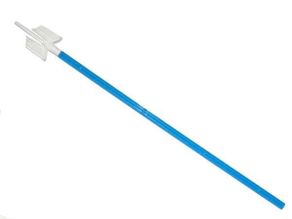

Vial with preservative fluid
Picture taken from: http://www.labnews.co.uk/cms_images/Image/Prod-Dec-07/31-NOV.jpg

Inside of the ThinPrep Processor
Picture taken from: http://cyto.igabinet.pl/data/user_files/image/TP%20Processor%202.JPG
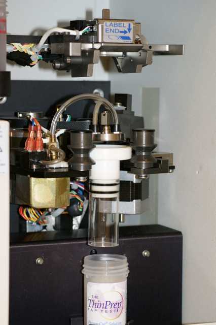

Slide produced from ThinPrep
Picture taken from: http://www.muliabrothers.com/Picture1TP.jpg

Difference in the quality of the slides produced by Conventional and ThinPrep method
Picture taken from: http://imaginis.com/graphics/cervical-cancer/pap_smear.gif
An endobrush, spatula or broom-like brush will be used to collect the cervical cells. After which, the brush will be rinsed vigorously in a vial containing preservative fluid. The vial will then be loaded into the ThinPrep machine for processing.
A suction filter will be inserted into the vial to filter the cells, separating the cervical cells from any debris, blood, mucosal or inflammatory cells. The cervical cells will then be imprinted on the glass slide. After which, the machine will gently drop the slide into 95% xylene for fixing. After fixation, the slides will go through pap staining in the autostainer, Leica Autostainer XL. The slides will then be mounted and screened under the microscope for any abnormalities.
2) Processing of Non-gynaecological specimens
The processing of non-gynae specimens is quite different from gynae specimens.
First of all, the specimens will undergo cytospin to obtain the pellet. To the pellet, cytospin collection fluid (light green colour) will be added to the cells. The amount the fluid to be added depends on the size of the pellet, i.e the fluid added must be at least twice the amount of the size of the pellet. A pipette was then used to mix up the mixture.
After these, a cytofunnel chamber is prepared. A glass slide, together with a filter card are attached to the cytoclip and cytofunnel. The whole cytofunnel chamber is then loaded into the cytospin. Due to the pressure from the spinning motion in the cytospin, the cells will be imprinted in the slide, according to the size of the filter card. Similarly, the slides will be stained in an autostainer.
Although both gynae and non-gynae specimens are stained with the same stains, they are stained in different autostainers. This is to prevent contamination of the different types of specimen. After staining, the slides will be mounted and screened under the microscope for abnormalities.

Process of assembling the cytofunnel chamber
Picture taken from: http://www.thermo.com/eThermo/CMA/PDFs/Various/File_24579.pdf
In the cytology lab, all equipments used will be disinfected with reagent CIDEX OPA. For the other disposable equipments like pasteur pipette and supernatants, they will be disinfected and discarded into a small bin of dissolved chlorine tablet.